3D Printing Enhances Medical Imaging and Education
If a picture of a heart is worth a thousand words, imagine the value of holding it in your hand!
Cardiac imaging specialists at Temple University Hospital typically use advanced cardiac imaging techniques like 3D echocardiography and cardiac CT angiography to guide their colleagues in interventional cardiology, electrophysiology and cardiac surgery. In essence, they provide patientspecific road maps for cardiac procedures. Though imaging technology has improved over the years, tangible 3D-printed anatomic models add a new dimension.
“3D printing is an incredible advance in cardiac imaging,” says Pravin Patil, MD, a non-invasive cardiologist in the Temple Heart & Vascular Institute who specializes in advanced cardiac imaging. “What better way to understand your patient’s cardiac anatomy than to hold it in your hand?”
Dr. Patil has recently taken advantage of a new 3D printer housed in the Health Sciences medical library to print models of cardiac patients’ hearts using their CT imaging studies.
“After the patient gets a cardiac CT scan, we extract the 3D anatomical structure of clinical interest using special software and generate a file containing the 3D data,” Dr. Patil says. “The
file is then sent to the printer, where the 3D model is reconstructed using a special kind of plastic filament and additive layer manufacturing techniques.”
The patient-specific plastic 3D anatomic models help cardiologists visualize and understand their patient’s anatomy; they can then perform test interactions with prostheses and plan procedures before they work on the patient’s actual heart.
“A perfect example is the left atrial appendage exclusion procedure – a procedure for patients who have atrial fibrillation and contraindications to long-term blood thinners,” Dr. Patil says. “In planning for that procedure, these patients often get cardiac CT scans, but now we can also print a 3D model of their heart. When I review the CT images with the heart team prior to the procedure, I can also hand them a model of that patient’s heart including the left atrial appendage; they can actually test different sizes of prostheses in the model to see how well they fit.”
The models don’t yet share the same material properties of human tissue. They are currently printed in a plastic that is stiffer than human tissue but still offers significant advantages over traditional imaging techniques. “The models give us an idea of whether a procedure is technically feasible and what the potential challenges will be during the procedure,” Dr. Patil says. “It’s all part of making procedures successful and safer.”
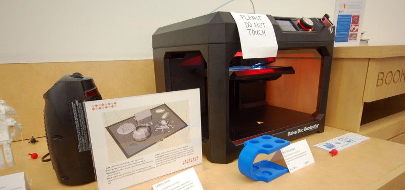
This is just one of the many ways the MakerBot™ Replicator printer is currently being used at Temple and, according to library staff, one of the nearly limitless applications for 3D printing in medicine.
“The main objective of the 3D printer is to support the mission of the Health Sciences Campus, so for us, objects related to patient care, education, or research have the highest priority,” says Barbara Kuchan, Director of the Health Sciences Libraries. “We’re offering this technology to faculty members and students throughout the entire Health Sciences Campus, not just the medical school, although we’ve seen it used frequently in that capacity.”
The MakerBot Replicator uses the extrusion method of printing, which involves laying down one layer at a time of a plastic called polylactic acid (PLA) filament. The printing process builds models from the base upward and usually takes several hours to complete, depending on the size of the model.
To introduce the new printer, library staff offered free 3D printing during the first week of February.
“We had quite a number of requests – so many that we had to cut it off a little early,” Kuchan says. “Some were personal items, but we also had requests from research labs for tools and devices and students who wanted anatomical models for studying. People have been really inventive with the things they have created, which makes it exciting.”
Down the road, the library hopes to purchase another printer that will allow objects to be printed in two different colors or materials. Some printers also use dissolvable supports to help printed objects better keep their shape.
“The technology is only going to improve, so we hope to have more options available in the near future,” Kuchan says.
The librarians plan to offer workshops on how to use the printer and how it might be used in various fields. For Dr. Patil and his colleagues in the Heart & Vascular Institute, the technology has come at just the right time.
“It’s truly hands-on, which is invaluable to us as educators and physicians,” he says.